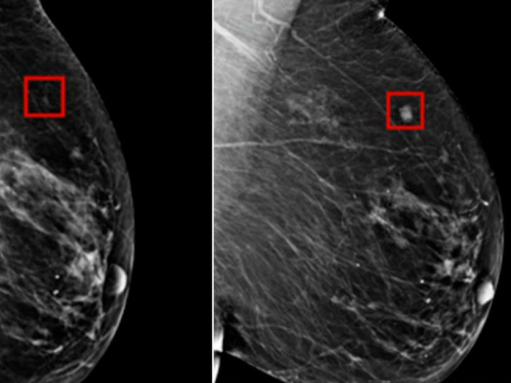
Your mammogram report says “dense breast tissue.” That phrase can feel confusing. Or worrying. Nearly half of women over 40 have dense breasts. Dense tissue can hide tumors on a standard mammogram. Radiologists call this “masking.” Dense tissue is also, on its own, a modest risk factor for breast cancer.
This article explains what dense breast tissue means. You’ll learn why MRI often catches what mammograms miss. And what to do next if you live near Norcross, Jonesboro, or Gainesville, GA.
Breast cancer caught early is easier to treat. That’s true for every woman. It matters even more when standard screening has blind spots.
Quick answer: If you have dense breast tissue, mammography alone may not be enough. Breast MRI offers significantly higher sensitivity for detecting cancer in dense tissue, especially for women at higher risk.
What Is Dense Breast Tissue?
Breast density compares glandular and connective tissue to fatty tissue. It has nothing to do with breast size or firmness. Radiologists grade density into four BI-RADS categories, using the same mammogram that screens for cancer.
- Category A: Almost entirely fatty tissue (about 10% of women)
- Category B: Scattered areas of dense tissue (about 40% of women)
- Category C: Heterogeneously dense tissue (about 40% of women)
- Category D: Extremely dense tissue (about 10% of women)
Categories C and D count as “dense breasts.” That’s nearly half of all women screened. Density tends to decrease with age. It also runs in families.
How Do I Know If I Have Dense Breasts?
Your mammogram report lists your BI-RADS density category. Georgia law requires facilities to tell you, too. It’s called Margie’s Law, named for a Savannah woman whose cancer was initially missed. The law took effect in 2019.
Why Dense Tissue Hides Tumors
A 1.5T open MRI still delivers accurate, diagnostic-quality images. It’s a dependable option for confirming ligament and cartilage injuries, and it remains a trusted standard across sports medicine.
Open MRI for Claustrophobic Athletes
Traditional closed MRI tubes feel tight. Open MRI removes that barrier. Athletes get a clear scan without the closed-in feeling, and parents can often stay close during a young athlete’s exam.
The Data Behind MRI’s Advantage
Picture searching for a snowball buried in snow. That’s mammography in dense breast tissue. Now picture thermal imaging finding the same snowball by its heat. That’s closer to how MRI works. It highlights blood flow feeding a tumor, not just its shape. Cancers typically recruit new blood vessels. MRI contrast dye makes that activity visible.
Published research backs this up. A 2020 study in JAMA compared abbreviated breast MRI to 3D mammography (tomosynthesis) in women with dense breasts and average risk.
| Screening Method | Sensitivity for Invasive Cancer | Detection Rate (per 1,000 women) |
| Breast MRI | 95.7% | 11.8 |
| 3D Mammography | 39.1% | 4.8 |
Sensitivity for Detecting Invasive Cancer in Dense Breasts
Abbreviated breast MRI vs. 3D mammography, women with dense breast tissue
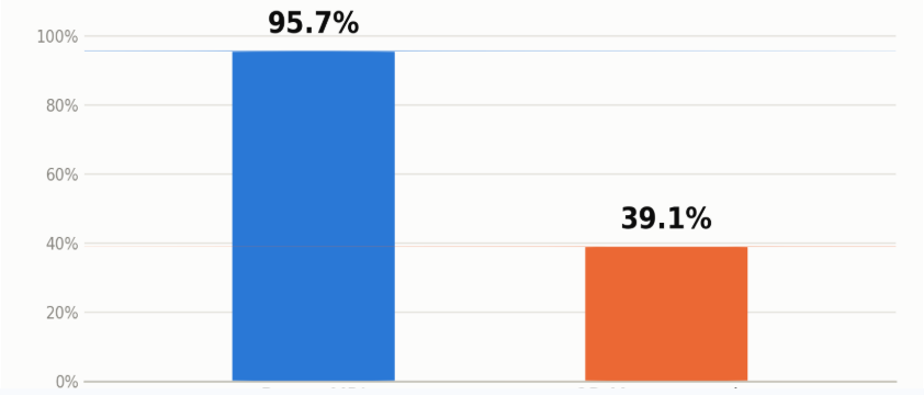
Source: Comstock et al., JAMA 2020 (abbreviated MRI vs. digital breast tomosynthesis, dense-breast screening study)
MRI found significantly more cancers than mammography alone. Especially invasive cancers, in dense tissue. Fewer cancers get missed between annual screenings, too.
The American College of Radiology recommends supplemental MRI for women with extremely dense breasts or added risk factors. At MRI Imaging Specialist, board-certified radiologists Dr. Thomas Turek and Dr. Marzieh Thurber apply that guidance to build individualized screening plans. Dr. Thurber holds memberships in the Society of Breast Imaging and the National Consortium of Breast Centers, and has extensive experience with 3D tomosynthesis and breast MRI.
What To Do If You Have Dense Breasts
- Ask for your BI-RADS category. Request it after every mammogram. It’s your right under Georgia law.
- Review your personal risk factors. Family history matters. So does a prior biopsy. Your provider can calculate your lifetime risk.
- Talk with your provider about supplemental screening. MRI is often recommended for category C or D, especially alongside other risk factors like family history or a genetic mutation.
- Schedule your breast MRI. Same-day scheduling is available at MRI Imaging Specialist, with results read by a radiologist quickly.
- Bring your prior imaging. This helps radiologists compare results accurately, year over year.
Tip: Dense breast tissue is common. It’s not a diagnosis. But it does change your screening plan
Take the Next Step Toward Better Detection
Dense breast tissue doesn’t mean something is wrong. It means mammography alone may not tell the full story. Breast MRI adds clarity. It finds cancers earlier, when treatment works best and options stay broader. MRI Imaging Specialist serves Norcross, Jonesboro, and Gainesville, GA. Our board-certified radiologists read every scan personally, with same-day and STAT turnaround available.
Schedule your breast MRI today. Visit our Women’s Imaging Services page, or call the location nearest you.
Request an AppointmentNorcross
(678) 969-0904
6760 Jimmy Carter Blvd, Suite 165
Jonesboro
(678) 545-6778
6568 Tara Blvd, Suite B
Gainesville
(678) 989-4566
955-E Interstate Ridge Business Park