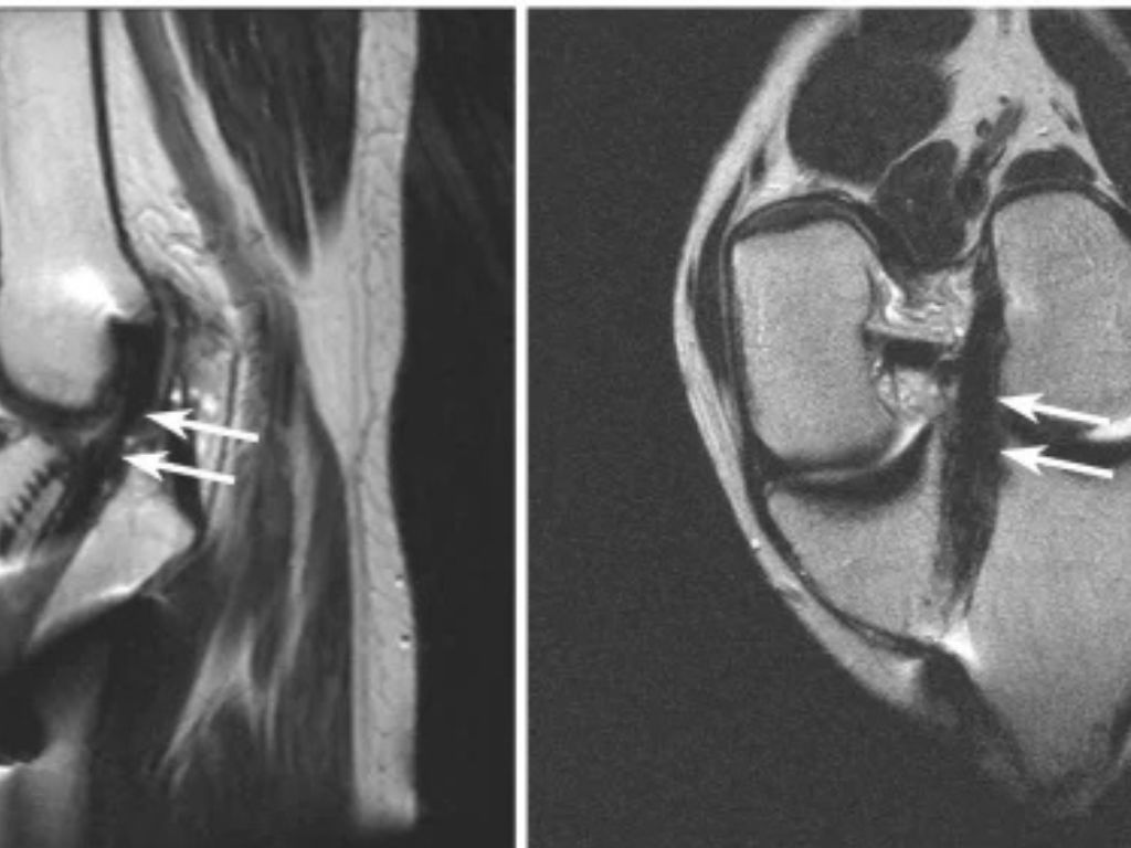
A twisted knee can end a season. One “pop” often means something is torn. Athletes want answers fast. Waiting for a diagnosis is stressful. Swelling hides the real damage. Instability makes every step feel unsafe. A physical exam alone can’t confirm a torn ACL.
This case study shows how a sports injury MRI found a torn ACL fast, and got one athlete into surgery on time. You’ll see the imaging process, the diagnosis, and the exact next steps to take after a knee injury.
Quick Answer: A sports injury MRI uses magnetic fields, not radiation, to capture detailed images of ligaments, tendons, and cartilage. It confirms an ACL tear with high accuracy, often the same day, so your doctor can plan treatment fast.
What Is a Sports Injury MRI?
A sports injury MRI scans the soft tissue around a joint. It shows what an X-ray cannot: ligaments, tendons, cartilage, and muscle. That detail matters most in the first days after an injury, when swelling can make a hands-on exam unreliable.
For a suspected torn ACL, MRI can reveal:
- Ligament tears, partial or complete
- Meniscus damage
- Bone bruises
- Cartilage injuries
- Tendon strains
Different MRI systems suit different patients and injuries. MRI Imaging Specialist offers three options, so your scan matches your body and your injury.
Who needs a sports injury MRI? Anyone with knee swelling, instability, or a “pop” sound at the time of injury should ask their doctor about one. The same goes for shoulder, ankle, and wrist injuries that don’t improve after a few days of rest.
3T MRI Open Bore HD for Sports Injuries
A 3T MRI captures sharper detail than standard scanners. Imaging research shows 3T systems can be highly accurate for partial ACL tears, where subtle fiber damage is easy to miss on lower-field imaging. For competitive athletes, that extra clarity can shape the entire treatment plan.
High Field 1.5T Open MRI
A 1.5T open MRI still delivers accurate, diagnostic-quality images. It’s a dependable option for confirming ligament and cartilage injuries, and it remains a trusted standard across sports medicine.
Open MRI for Claustrophobic Athletes
Traditional closed MRI tubes feel tight. Open MRI removes that barrier. Athletes get a clear scan without the closed-in feeling, and parents can often stay close during a young athlete’s exam.
Real-World Case: Diagnosing a Torn ACL
Case Snapshot: This composite scenario reflects a common patient experience at MRI Imaging Specialist. A 17-year-old soccer player felt her knee buckle during a game. She heard a pop. Swelling followed within hours, and by the next morning her knee felt unstable.
She couldn’t bear weight the next day. Her physician suspected a ligament injury but needed imaging to confirm it and rule out other damage before deciding on treatment. The table and timeline below show how the imaging options compare and how the case moved from injury to a surgical plan.
| Feature | 3T MRI Open Bore HD | 1.5T High Field Open MRI | Open MRI |
| Best for | Fine detail, partial tears | General ligament & cartilage imaging | Claustrophobia-friendly imaging |
| Partial ACL tear accuracy | Up to ~95% | ~25–53% (varies by study) | Varies by field strength |
| Scan experience | 70 cm open bore, faster detail capture | Open-style comfort | Most spacious, open design |
| Great for | Competitive athletes needing precision | Most sports injury cases | Patients uneasy in enclosed spaces |
*Based on published imaging literature; individual accuracy varies by scanner, protocol, and injury type.
Illustrative Case Timeline: Injury to Surgery
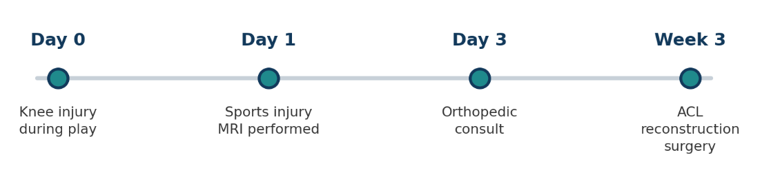
The scan confirmed a complete ACL tear. It also showed early meniscus involvement, a detail the physical exam missed. Her orthopedic surgeon used the MRI images to plan reconstruction surgery within weeks of the injury, rather than guessing based on symptoms alone.
Early, accurate imaging matters. MRI Imaging Specialist’s radiology team, led by board-certified radiologists Dr. Thomas Turek and Dr. Marzieh Thurber, brings more than 50 years of combined experience to every sports injury scan. Reports are typically ready the same day, with STAT reads available in about two hours, so patients and physicians aren’t left waiting on answers.
Step-by-Step: What To Do After a Sports Injury
- Stop playing immediately. Don’t push through knee pain or try to “walk it off.”
- Apply R.I.C.E. Rest, ice, compression, and elevation reduce swelling fast.
- See a doctor within 24 to 48 hours. Early evaluation catches complications sooner.
- Get a sports injury MRI. Confirm the diagnosis before choosing a treatment path.
- Follow your treatment plan. Start physical therapy or surgery prep as soon as your doctor clears you.
⚠️Warning: A knee that buckles or gives out may signal a torn ACL. Don’t wait to get it scanned.
Get Answers Fast With Sports Injury MRI
A fast, accurate diagnosis changes everything. It shortens the wait between injury and treatment. It clears the path to surgery or recovery. Most importantly, it gets athletes back to their sport with a real plan instead of guesswork.
MRI Imaging Specialist offers 3T MRI Open Bore HD, 1.5T High Field Open MRI, and open MRI across Norcross, Jonesboro, and Gainesville, GA. Same-day and STAT reads mean faster answers for urgent sports injuries, and your referring physician receives results quickly enough to act on them.
Bring your physician’s order, a photo ID, and a list of any prior imaging when you arrive. Scans typically take 30 to 45 minutes, and technologists explain each step beforehand so nothing feels rushed. Don’t wait on knee pain or a suspected ACL tear. Schedule your sports injury MRI today and get the clarity your treatment plan needs.
Request an AppointmentNorcross
(678) 969-0904
6760 Jimmy Carter Blvd, Suite 165
Jonesboro
(678) 545-6778
6568 Tara Blvd, Suite B
Gainesville
(678) 989-4566
955-E Interstate Ridge Business Park