That headache won’t quit. It’s been three weeks.
Now the pain spreads behind your eyes, and nothing helps.
A quick search turns up one scary word: tumor.
Suddenly you need real answers, not guesses.
Most headaches have nothing to do with a tumor. But some symptoms are different. They linger, worsen, or bring new problems like blurred vision or numbness. This guide explains exactly when a brain tumor MRI is needed. You’ll learn the warning signs, how the scan works, and what to do next.
Quick Answer: A brain MRI is recommended when headaches are new, worsening, or paired with vision changes, weakness, seizures, or persistent dizziness. MRI remains the clearest way to rule a tumor in or out, without guesswork.
What Is a Brain Tumor MRI?
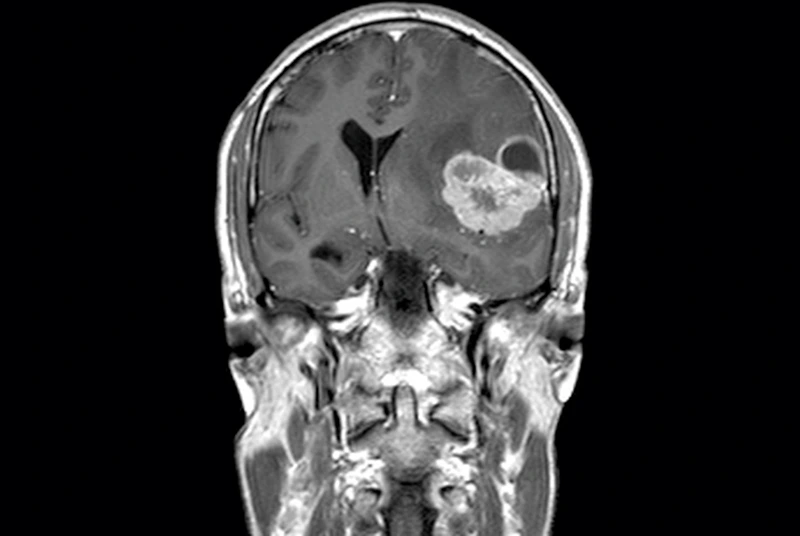
A brain tumor MRI uses magnetic fields and radio waves, not X-rays. It builds detailed cross-section images of brain tissue from multiple angles. Radiologists use these images to spot abnormal growths, swelling, or shifts in brain structure that a physical exam alone cannot reveal. The scan is painless and non-invasive. Most patients simply lie still while the machine captures images, and results typically reach your doctor within a day or two.
- Magnetic field and radio waves power the imaging, not radiation.
- Cross-sectional images are captured from multiple angles.
- Contrast dye, when used, highlights blood flow and tumor borders.
- Scan time typically runs thirty to forty-five minutes.
- A board-certified radiologist reads and reports on every image.
Who Should Consider a Brain Tumor MRI
Certain factors raise the case for imaging. New headaches after age fifty deserve attention. A personal or family history of cancer matters too. Weakened immune systems raise risk. Anyone with sudden neurological changes, like slurred speech or facial drooping, should seek same-day evaluation, not a wait-and-see approach.
MRI vs. CT Scan for Brain Tumors
CT scans work fast. They spot bleeding or major structural damage well, which makes them useful in emergencies. MRI shows far more detail in soft tissue. That extra clarity makes MRI the preferred test whenever a brain tumor is suspected. Many doctors order both: a CT first for speed, then MRI for precision.
| Feature | MRI | CT Scan |
| Radiation exposure | None | Yes, low dose |
| Soft tissue detail | Excellent | Moderate |
| Typical scan time | 30–45 minutes | 5–10 minutes |
| Best for | Detailed tumor evaluation | Emergency bleeding, fast triage |
| Contrast option | Gadolinium-based | Iodine-based |
Brain Tumor MRI’s in Georgia: What Sets Us Apart
Not every imaging center offers the same turnaround or expertise. MRI Imaging Specialist has served Georgia patients for over thirty years as a family-owned practice. Board-certified radiologists, with over fifty years of combined experience, interpret every scan. Locations in Norcross, Jonesboro, and Gainesville keep quality imaging close to home, with same-day and STAT reading options available for urgent cases.
Real-World Proof: Why Early Imaging Matters
Consider a common scenario. A forty-two-year-old patient reports morning headaches for a month. Over-the-counter medicine stops helping. Vision blurs during long workdays, and she starts missing details at her job. Her primary care doctor orders an MRI to check for a structural cause. The scan rules out a tumor. It confirms a treatable sinus issue instead. Early imaging brought fast answers, real relief, and one less thing to worry about.
Real-World Proof: Why Early Imaging Matters
New or worsening headaches
Especially morning headaches or ones that wake you from sleep
Headaches with vomiting
Nausea or vomiting alongside head pain
Vision changes
Blurred, double, or partial vision loss
Persistent balance problems
Ongoing dizziness, stumbling, or coordination issues
New weakness or numbness
In an arm, leg, or one side of the face
Seizures
A first-time seizure with no prior history
Personality or memory changes
A first-time seizure with no prior history
Source: Compiled from Dana-Farber Cancer Institute, MD Anderson, and Cleveland Clinic patient guidance.
Board-certified radiologists note that most headaches are not caused by tumors. Still, new neurological symptoms deserve prompt imaging. An MRI gives clear answers fast, easing worry either way and guiding the next step in care.
Step-by-Step: What to Do If You Suspect a Brain Tumor
Follow these steps if new or worsening symptoms concern you.
- 📝 Track your symptoms. Note headache timing, triggers, and severity in a simple log.
- 🩺 See your doctor. Describe new or worsening symptoms clearly and completely.
- 📋 Ask about an MRI referral. Request imaging if any red flags apply to you.
- 📅 Schedule your scan. Same-day and STAT appointments are available at most locations.
- 📞 Review results with a radiologist. Get clear next steps and a plan right away.
⚠️ Seek emergency care immediately for a sudden severe headache, a first seizure, or loss of consciousness. Don’t wait for a scheduled appointment.
Get Your Brain Tumor MRI in Georgia Today
Waiting rarely helps and often adds stress. A brain tumor MRI brings clarity fast. It confirms what’s happening inside your head, one way or the other. Then you and your doctor decide on next steps together, backed by real imaging instead of worry. Patients across Norcross, Jonesboro, and Gainesville trust local, board-certified radiologists for fast, accurate answers.
Schedule your brain tumor MRI today at MRI Imaging Specialist. Visit our MRI imaging services page or call your nearest Georgia location below to book an appointment.
Request an AppointmentNorcross
(678) 969-0904
6760 Jimmy Carter Blvd, Suite 165
Jonesboro
(678) 545-6778
6568 Tara Blvd, Suite B
Gainesville
(678) 989-4566
955-E Interstate Ridge Business Park