He Couldn’t Form Words. An MRI Told His Doctors Exactly Why.
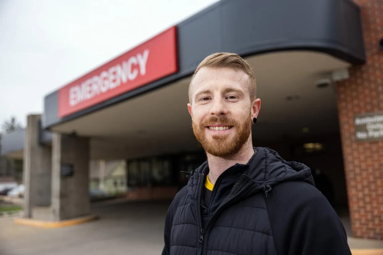
Nick Vance was 37 years old when he woke up one morning in November 2024 and tried to turn on his lights. He opened his mouth to say “Alexa, turn the lights on” — and what came out was word salad.
He called his mother. He could barely get out: “Help me. Can’t talk.“
His parents rushed him to the ER. Within hours, an MRI gave his medical team a precise answer: Nick had experienced an ischemic stroke — a blood clot blocking flow to the part of his brain responsible for speech. His story was documented by University of Iowa Health Care in May 2025. He made a full recovery.
The difference between full recovery and permanent disability in stroke care almost always comes down to two things: recognizing the warning signs early, and getting to the right imaging fast. For patients in Georgia, that imaging is available close to home.
What Is an Ischemic Stroke — and Why Every Minute Matters
An ischemic stroke occurs when a blood clot cuts off oxygen to part of the brain. Without oxygen, brain cells begin dying within minutes.
Neurologists at UCLA, publishing in the Stroke journal, quantified this precisely: during a typical large-vessel ischemic stroke, the brain loses 1.9 million neurons every minute untreated. Every hour without treatment, the brain ages 3.6 years.
The treatment window for the clot-dissolving drug tPA is just 3 to 4.5 hours from symptom onset. Every minute of delay narrows that window and deepens the damage.


Getting the right imaging, fast, is what makes treatment possible. For Georgia residents in Norcross, Jonesboro, or Gainesville, MRI Imaging Specialist offers brain MRI with same-day reads and STAT turnaround — because in stroke care, report timing is everything.
Recognizing the Warning Signs (F.A.S.T.)
Stroke symptoms can appear suddenly and without warning. The most widely used screening framework is the FAST acronym:

Other sudden symptoms that warrant immediate attention: severe unexplained headache, sudden vision problems, difficulty walking, or sudden dizziness.
One dangerous pattern: mild symptoms that seem to pass on their own. These may be transient ischemic attacks (TIAs) — “mini-strokes” — which are serious warning signs. According to the American Heart Association, 1 in 3 people who have a TIA will go on to have a full stroke, often within days or weeks.
If you’re cleared at the ER following a suspected TIA or stroke, follow-up brain MRI is often the recommended next step. The emergency team stabilizes — imaging specialists help answer what happened and what your risk looks like going forward.
Brain MRI in Georgia — MRI Imaging Specialist
For patients in the Norcross, Jonesboro, and Gainesville areas, MRI Imaging Specialist provides expert brain MRI with board-certified radiologists, same-day reads, and two-hour STAT turnaround for urgent cases.
Request an AppointmentNorcross
(678) 969-0904
6760 Jimmy Carter Blvd, Suite 165
Jonesboro
(678) 545-6778
6568 Tara Blvd, Suite B
Gainesville
(678) 989-4566
955-E Interstate Ridge Business Park
- Same-day and STAT reads.
- Same-day turnaround on standard reads
- two-hour turnaround on urgent reads — because in neurology, the report timing matters.
Board-certified radiologists. All imaging is read by credentialed specialists who understand the nuances of stroke imaging, DWI patterns, and vascular findings.
Flexible payment options. MRI Imaging Specialist works with insurance, and offers cash pricing, attorney liens, and worker’s compensation arrangements.
A physician’s referral or order is typically required for diagnostic brain MRI. If you’ve experienced symptoms that concerned you — or if your doctor has mentioned brain imaging and you’re not sure what to do next — the team at MRI Imaging Specialist can walk you through the process.
Why MRI Is the Critical Tool in Ischemic Stroke Diagnosis
When a patient arrives with stroke symptoms, the first imaging priority is distinguishing ischemic stroke (blocked vessel) from hemorrhagic stroke (bleeding in the brain) — because the treatments are opposite. Giving a clot-dissolving drug to someone who is actually bleeding can be fatal.
CT scans are fast at detecting bleeding, but for ischemic stroke they have a significant limitation. Research published in PMC shows that non-contrast CT has a sensitivity of only 40–60% for ischemic stroke in the first six hours — missing roughly half of all cases when it matters most.
MRI — specifically diffusion-weighted imaging (DWI) — detects the cellular changes that occur within minutes of a blood flow disruption, making ischemic damage visible long before it appears on CT. The same research confirms MRI is approximately five times more sensitive than CT for diagnosing acute ischemic stroke.

Source: PMC9399663 — MRI in Acute Ischemic Stroke; AHA Stroke Journal
Consider Kate, a 49-year-old documented by MedPark Hospital’s neurology team: she arrived unable to complete simple tasks. Her neurologist ordered an MRI with stroke protocol. The scan revealed extensive ischemia and a large vessel occlusion, enabling immediate mechanical thrombectomy. The clot was removed in a single procedure. She spent one night in the ICU.
What MRI Shows That CT Cannot
MRI for stroke is typically multimodal — several imaging sequences run together to give a complete clinical picture:
DWI
(Diffusion-Weighted Imaging) — Detects cytotoxic edema — the cellular swelling that begins within minutes of a blockage. This is what catches strokes early.
DWI
Fluid-Attenuated Inversion Recovery) — Helps distinguish how old a stroke is. DWI positive + FLAIR negative = stroke within the last 4.5 hours, critical for tPA eligibility. Basis of the landmarkWAKE-UP trial.
MRA
MR Angiography visualizes the blood vessels themselves, identifying the location and extent of blockages — essential for planning thrombectomy.
PERFUSION
Maps blood flow across the brain in real time, distinguishing dead tissue from salvageable tissue — guiding decisions about whether intervention is still worthwhile.
Together, these sequences give physicians a complete map of what happened, where, how long ago, and what can still be done.
A 2024 study in Radiology found that AI-accelerated MRI can now complete the full diagnostic protocol in just over 3 minutes — making the “MRI takes too long” argument increasingly obsolete.
The Bottom Line
Nick Vance couldn’t speak. An MRI told his doctors exactly where in his brain the damage was, what caused it, and what to do about it. He made a complete recovery.
Stroke is time-sensitive in a way few emergencies are. Every minute of delay is measurable brain loss — and every minute of fast, accurate diagnosis is brain saved.
If you recognize the F.A.S.T. signs, call 911 first. If you’re in Georgia and need follow-up neurological imaging, expert brain MRI with board-certified radiologists is available close to home at MRI Imaging Specialist — in Norcross, Jonesboro, and Gainesville.
This article is for informational purposes only and does not constitute medical advice. If you believe you or someone around you is having a stroke, call 911 immediately. Please consult your physician or a qualified healthcare provider regarding your individual health situation and any imaging or treatment decisions.


