A blurry spot on a brain scan can change everything. Numbness. Vision trouble. Fatigue that won’t quit. Your doctor mentioned multiple sclerosis, and now you’re searching for answers.
You’re not alone. Nearly 1 million Americans live with MS. Most got their answer from one test: an MRI. This guide explains how a multiple sclerosis MRI works, what recent research changed, and what to expect at your scan.
Quick Answer: Yes. MRI is the most reliable tool for detecting multiple sclerosis. It reveals lesions in the brain, spinal cord, and optic nerve that signal active MS damage. No blood test can do this alone.
What Is an MRI’s Role in Diagnosing MS?
MS attacks myelin, the protective coating around nerve fibers. That damage leaves scars called lesions. MRI is the only tool that shows these lesions clearly enough to confirm a diagnosis.
Neurologists rely on MRI to:
- Spot lesions in the brain and spinal cord
- Track new or growing damage over time
- Rule out conditions that mimic MS
- Excess body weight
- Confirm a diagnosis under the McDonald Criteria
What Do MS Lesions Look Like on an MRI?
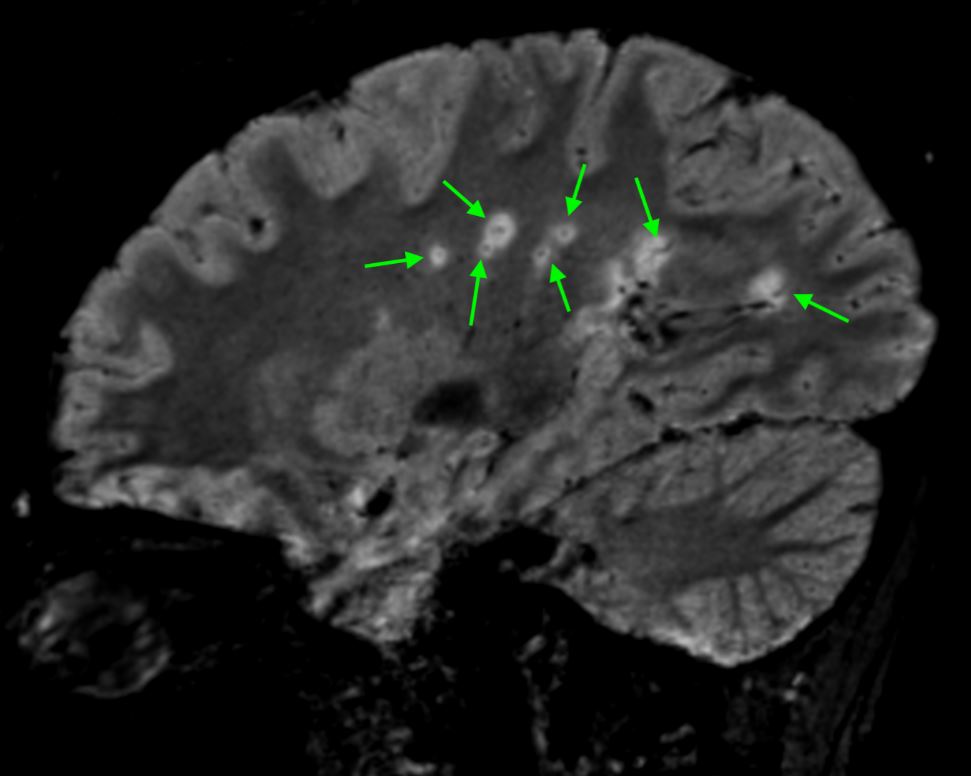
MS lesions show up as bright spots on T2-weighted MRI scans. Radiologists check five specific regions: periventricular, cortical or juxtacortical, infratentorial, spinal cord, and the optic nerve.
Two newer markers add precision:
- Central vein sign. A thin vein runs through the center of true MS lesions. This feature helps rule out look-alike conditions, like small vessel disease.
- Paramagnetic rim lesions. A dark iron ring around a lesion signals chronic, ongoing inflammation.
Brain MRI vs. Spinal MRI for Multiple Sclerosis
Brain MRI catches most MS lesions. Spinal MRI catches the rest, especially in patients with normal-looking brain scans but real symptoms. Doctors often order both for a complete picture.
Contrast dye plays a key role too. Active lesions absorb gadolinium and light up on the scan. Older, inactive lesions don’t. This distinction helps your doctor tell whether MS is currently active or stable.
Real-World Proof: How Recent Research Changed MS Diagnosis
MS diagnosis just got more precise. In 2024, the McDonald Criteria, the diagnostic standard neurologists use worldwide, were revised for the first time since 2017.
Herniated Disc Symptoms in Georgia Patients
Patients across Georgia report similar warning signs:
- Sharp or burning lower back pain
- Pain that radiates into the hip, leg, or foot
- Numbness or tingling in the leg or foot
- Muscle weakness affecting balance or grip
- Pain that worsens when sitting, coughing, or bending
Real-World Proof: How MRI Confirms a Herniated Disc
| Diagnostic Factor | 2017 Criteria | 2024 Criteria |
| Topographic regions checked | 4 regions | 5 regions (adds optic nerve) |
| Central vein sign | Not included | Supportive evidence for MS |
| Paramagnetic rim lesions | Not included | Supportive evidence for MS |
CSF biomarkers | Kappa free light chains not included | Added as substitute evidence |
| Radiologically isolated syndrome | Not classified as MS | Can qualify as MS in specific cases |
According to a 2025 guide for radiologists published in the American Journal of Roentgenology, these updates were built to catch MS earlier and reduce misdiagnosis, a persistent problem in the field. Earlier detection means earlier treatment, and earlier treatment means better long-term outcomes.
For patients, this means fewer inconclusive scans and fewer years spent waiting for a clear answer. Research is also moving toward AI-assisted MRI analysis, which combines imaging with blood biomarkers to flag disease patterns radiologists might otherwise miss.
Common MS Lesion Sites on MRI (2024 McDonald Criteria: 5 Topographic Regions)
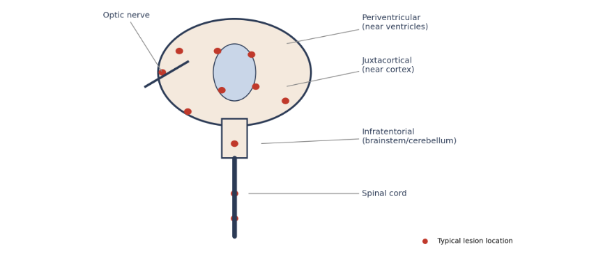
Common MS lesion sites on MRI under the 2024 McDonald Criteria’s five topographic regions.
What to Expect During Your MS MRI
Getting scanned is simpler than most patients expect.
- Schedule your appointment. Call your nearest location to book.
- Complete a safety screening. Staff check for metal implants or pacemakers.
- Change into a gown. Remove jewelry and anything metal.
- Receive contrast dye, if ordered. Gadolinium highlights active inflammation.
- Lie still during imaging. The scan itself takes 30 to 60 minutes.
- Review results with your doctor. A board-certified radiologist reads your scan, usually the same day, and sends the report to your doctor.
⚠️Tip: Feeling claustrophobic? Tell the technologist beforehand. Our staff can walk you through options to help you feel comfortable.
Get Clarity With an MS MRI Near You
You don’t have to wait and wonder. A multiple sclerosis MRI gives you real answers, backed by the latest diagnostic research.
MRI Imaging Specialist offers advanced MRI imaging at three convenient Georgia locations: Norcross, Jonesboro, and Gainesville.
We also offer CT/CT angiography, digital X-ray, 3D/4D ultrasound, I-CAT 3D imaging, mammography, bone density scans, and women’s imaging services.
Ready for answers? Schedule your MRI today at your nearest location.
Request an AppointmentNorcross
(678) 969-0904
6760 Jimmy Carter Blvd, Suite 165
Jonesboro
(678) 545-6778
6568 Tara Blvd, Suite B
Gainesville
(678) 989-4566
955-E Interstate Ridge Business Park